|
Chapter 2 : Physiology of the HeartChapter 2: Anatomy And Physiology Of The HeartThe heart is a hollow and complex organ which has an intricate system of 4 chambers pumping blood throughout the body. The upper chambers are the atria and the lower chambers are called the ventricles; the chambers are separated by a thin muscular wall called the septum. The right atria and right ventricle push deoxygenated from the body to the lungs to become oxygenated. While the left atrium and left ventricle move the oxygen-rich blood from the lungs to the rest of the body. Chapter 2A Anatomy of the Heart Video:The heart muscle cells generate, conduct and respond to electrical impulses spontaneously. Cardiac contraction is stimulated by impulses from the Sinoatrial node or SA node, which is the pacemaker of the heart, located in the right atrium. Once the SA node stimulates the impulse it travels between the atrium and ventricles, where the Atrioventricular node or AV node is located. Here the impulse stays for a few seconds before it travels to the purkinje fibers and these fibers trigger the ventricles to contract. The electrical activity of the heart is graphed in a wave pattern on an electrocardiogram (ECG/EKG). The electrocardiogram is a test that checks the conduction of the heart and any problems associated with it. It helps locate the cause of chest pain which may be due to inflammation, heart attack or angina. It can help find the cause of symptoms of heart diseases as well as show if medications are working effectively. To understand the pathology on an ECG it is important to know how a normal sinus rhythm works. Chapter 2B Physiology of the Heart Video:As the heart conducts, one heartbeat is a group of waves called P-QRS-T deflections. The P wave is the electrical activity of the atria. When the atrium is stimulated, the impulse is recorded as a P wave. The QRS-T complex shows the electrical activity of the ventricles. The QRS complex shows the depolarization of the ventricles and the T wave shows the repolarization of the ventricles. The PR interval shows the time the impulse takes to travel from SA node to AV node.
Learning Outcomes:You have completed Course I. Now you should be able to:


|
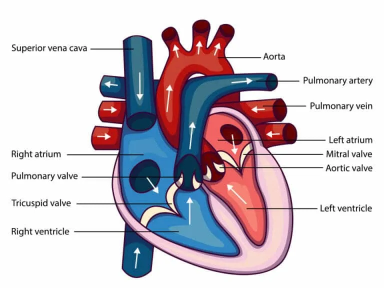
The heart is a hollow and complex organ which has an intricate system of 4 chambers pumping blood throughout the body. The upper chambers are the atria and the lower chambers are called the ventricles; the chambers are separated by a thin muscular wall called the septum. The right atria and right ventricle push deoxygenated from the body to the lungs to become oxygenated. While the left atrium and left ventricle move the oxygen-rich blood from the lungs to the rest of the body.
The heart muscle cells generate, conduct and respond to electrical impulses spontaneously. Cardiac contraction is stimulated by impulses from the Sinoatrial node or SA node, which is the pacemaker of the heart, located in the right atrium. Once the SA node stimulates the impulse it travels between the atrium and ventricles, where the Atrioventricular node or AV node is located. Here the impulse stays for a few seconds before it travels to the purkinje fibers and these fibers trigger the ventricles to contract. The electrical activity of the heart is graphed in a wave pattern on an electrocardiogram (ECG/EKG). The electrocardiogram is a test that checks the conduction of the heart and any problems associated with it. It helps locate the cause of chest pain which may be due to inflammation, heart attack or angina. It can help find the cause of symptoms of heart diseases as well as show if medications are working effectively. To understand the pathology on an ECG it is important to know how a normal sinus rhythm works.
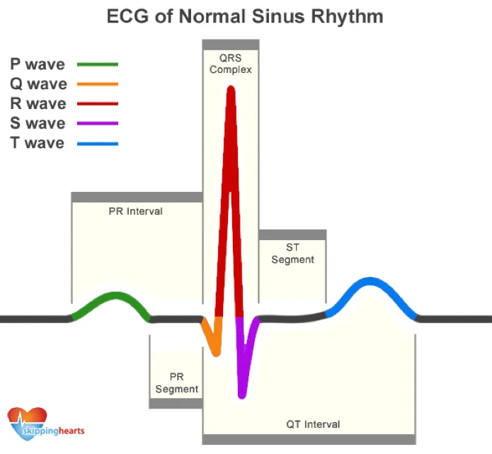
As the heart conducts, one heartbeat is a group of waves called P-QRS-T deflections. The P wave is the electrical activity of the atria. When the atrium is stimulated, the impulse is recorded as a P wave. The QRS-T complex shows the electrical activity of the ventricles. The QRS complex shows the depolarization of the ventricles and the T wave shows the repolarization of the ventricles. The PR interval shows the time the impulse takes to travel from SA node to AV node.
You have completed Course I. Now you should be able to: